
author: Mara Krause, 23.07.2026
A man, once having a perfectly normal life, now cannot move properly, cannot communicate with his loved ones, and requires around-the-clock caregiver support. This, unfortunately, is not an isolated case, but describes the life of many people with incurable brain diseases like Parkinson‘s, Alzheimer‘s or others.
Why incurable? Because the brain is almost hopelessly complex and medical science is not advanced enough yet.
„Flywire“ is an international project that started in mid-2020 with the goal of getting one step closer to understanding this complex biological computer sitting inside every one of our skulls. A Princeton-led team of scientists built a roadmap of an adult fruit fly (Drosophila melanogaster) neuron-by-neuron and synapse-by-synapse. “Any brain that we truly understand tells us something about all brains,” said Sebastian Seung, Princeton’s Evnin Professor in Neuroscience and a professor of computer science.
But that is not all: the scientists uploaded this map of the brain into a software and received a simulation of a virtual fruit fly. The computer model managed to mimic how a real fly processes taste and touch. However, the map and the simulation are not the same piece of work and they have not been done by the same team.
I am truly amazed by how science could create a digital animal brain. It can make predictions about its behaviour and shows what is possible when scientists collaborate. Whether this ever contributes to collecting more knowledge about the human brain and helping to cure diseases is a much longer road. But right now we can only simulate the fly’s brain.
Here is the more detailed, step-by-step road through making this digital fly. I read the papers several times and did some research to present the procedure here with a clear and hopefully retraceable structure.
How did they make the digital twin of the fruit fly?
On my first read of the paper, I was thrown into cold water because of all those specialised terms I didn’t get.MN9, G2N-1, bract 1 or 2, Zorro…
But after carefully researching those terms, the paper felt decoded. So, I divided the whole procedure into two steps: making the digital brain and then testing it.
making the digital brain
This step involves making the wiring diagram with every neuron and synapse, also called the connectome, then figuring out the neurotransmitters and finally developing a mathematical model (LIF) to give the computer program.
- Connectome
The connectome is a diagram of about 140,000 neurons and roughly 50 million synapses of the Drosophila melanogaster’s brain, although they used the central brain with more than 125,000 neurons in the paper. But the team actually didn’t map them alone, but researchers from around the world helped which makes this project so special.
To map the vast number of neurons and connections, they accessed the data from Flywire: an online platform and community project where a large community of researchers share their findings. This brought together 21 million electron-microscopy images which an AI used to make the connectome.
However, the AI that created the three-dimensional map was not accurate enough. Gamers, students and researchers around the world worked as proofreaders and assembled the pieces into one coherent whole. A brain map finished by volunteers was not what I expected to find behind a Nature paper but shows how much collaboration can achieve.
The scientific procedure: they used Electron Microscopy (EM) to find out about the neurons and synapses. They sliced a fly’s brain into incredibly thin layers and via an electron beam received images. Here a short aside for those interested in EM:
In EM, an electron beam is fired on brain slices. The electrons are scattered, absorbed or transmitted by the brain slice depending on density and material characteristics. The electrons are detected and a computer system turns the results into very detailed images. The computer receives millions of 2D images and with help of AI synthesises them to a 3D volume map.
2. Neurotransmitters
Knowing where the neurons are and how they are connected is good but not enough: neurotransmitters are the body’s chemical messengers. These molecules transmit signals between neurons and are as important as instruments in an orchestra.
In collaboration with other scientists and, of course, with AI, the research team was able to predict the neurotransmitters. AI looked at shape and details of synapses in electron microscope images to guess which chemical was likely being used.
Across the fly’s brain, about 55% of neurons are predicted to be cholinergic, 24% glutamatergic and 14% GABAergic. The remaining 7% are dopaminergic, octopaminergic or serotonergic. But what exactly does that mean?
For the simulation these predictions had to be reduced to a single question: does this neuron push the next one towards firing, or hold it back? Cholinergic neurons were treated as excitatory, and both GABAergic and glutamatergic ones as inhibitory. Excitatory neurons push next cells electrical charge higher and inhibitory neurons pull charge down
3. Leaky Integrate and -Fire (LIF)
The next step is to make this brain map work by developing a mathematical system that defines how digital cells should behave. For this project, they used the Leaky Integrate and -Fire (LIF) model.
This math determines how a cell builds up electrical charge from its neighbours (Integrate),
fires when hitting a threshold from the sum of signals it receives, then the electrical pulse to next cell (Fire),
and eventually lets the signal fade away if the action potential is too low (Leaky).
4. Spiking Neural Network Simulation
A computer finally calculates the electrical charge of every neuron based on the connections in the connectome. A visual simulation of this represents the digital fly.
testing the digital brain
After making the digital fly, the researchers needed to test their achievement, of course. The testing involved several tests, including the sense of taste, comparing it to real flies, testing leg movement and other tests. However, it seems to me that simulating the sense of taste was the test with most focus on, so I will go deeper into that.
- Simulating sense of taste
What matters more to a fly than eating? The team used the feeding circuit as one of their first real tests: the whole path from a leg touching sugar to the mouth reaching out for it.
Their goal was to verify that the computer model can send the signal from the taste-sensing neurons all the way to the motor neurons of the fly’s mouth, same as in a real fly.
It starts with the Gustatory Receptor Neurons (GRNs) on the fly’s leg and mouth that sense taste. The researchers looked at four kinds of taste: sugar, water, bitter, and a fourth type marked by the receptor Ir94e. With the LIF model they could simulate the whole chain of activated neurons.
The testing came down to switching on the sugar GRNs at a realistic firing rate and letting the signal travel through the network on its own to see where it lands. This network involves several layers and many cells as you can see in the visualisation below. Sugar activation landed on the Motor Neuron 9 (MN9), one of 16 motor neurons that move the proboscis, the tube-like mouthpart. MN9 lifts the rostrum, its largest segment, when the fly extends its mouthparts towards food.
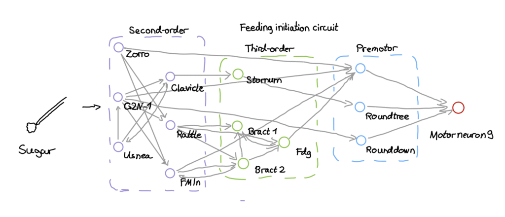
If activating sugar GRNs made MN9 fire, the model had reproduced the feeding reflex from wiring alone. And it did.
2. Comparison with real fly via optogenetics
To ensure the computer model is correct, the researchers compared the model to real living flies. One way to do this is via optogenetics. I first wondered if they really caught a fly, glued it to their experimental setup and somehow observed the firing in the fly. As abstract as this might sound, what they actually did wasn‘t far from my imagination.
They mounted the fly onto a glass slide with nail polish to keep it still. Then, a fascinating process enabled them a glimpse into the fly‘s brain: optogenetics.
They genetically modified the flies with a specific protein called CsChrimson in specific brain cells. This protein is light sensitive and when flashing a laser at the fly‘s head, they activated the cell.
If the computer predicted that a specific cell type would make the fly eat, they checked whether the real fly extended its proboscis when the laser hit that cell.
A different approach was inserting a protein that makes cells glow green when active. They gave the fly real sugar or water and used a microscope to see if the same cells lit up.
What did they achieve?
The comparison described above showed a remarkable accuracy of the model. Across 164 different tests, the model‘s predictions were 91% of the time correct
Another big achievement is that they understood taste interactions and the neural network of the taste circuit. But they not only understood it, they programmed a complete computer program that shows a digital fruit fly. In their paper they say themselves: „We find that the model makes predictions consistent with our empirical observations“
“How the brain functions depends critically on which neurons connect to which other neurons and the strength of their connections,” said Murthy, Princeton’s Karol and Marnie Marcin ’96 Professor of Neuroscience. “To have a full wiring diagram of the fly brain — as a Drosophila neurobiologist, this is something I’ve dreamed of since I started my lab in 2010.”
But the result that actually convinced me was Ir94e. Earlier work wasn’t sure what it did, and some assumed it was attractive. The model predicted the opposite: Ir94e should suppress extending the mouth towards it, like bitter. When the researchers checked this in real flies, the prediction held. That is what makes the model worth trusting. It didn’t just repeat what was already known, it called something new and got it right.
Which struggles and limitations did they face?
The team actually faced several struggles and limitations because they tried to simulate a biological organ with maths.
A huge limitation we have to consider is that the model is simplified and ignores complex physical shape of neurons (morphology) or gap junctions (short circuits between cells).
Another big mathematical problem was the basal Firing Rate. In the model, every neuron is silent (0Hz) unless stimulated, but real brains often have a background hum with activity even if nothing is happening.
Additionally, the model struggled with more complex chemicals like dopamine or serotonin because they don’t act like an on/off switch. The LIF model wasn’t designed for those signals.
Recently, it was discovered that the entire 3D brain map was actually left-right inverted and they had to carefully adjust their findings.
To be honest, the term digital twin might be a little exaggerated because it is not an actual twin and we still deal with many uncertainties.
What are hopes for the future?
From my side, hopes are high. But also, the researchers already have plans to improve their model and are rather optimistic to get one step closer to unravel the mysterious biological computer in our skulls. They want to include the physical shape of neurons (morphology), gather more details about receptors and use the model to study other behaviour like hunger, thirst or memory.
For many it might not sound too relatable to have mapped a fruit fly‘s brain because, well, it is still just a fruit fly. And while it does not have exactly the same neurons as humans, they are very similar and they actually have the same neurotransmitters like glutamate, acetylcholine, dopamine… The step from fruit fly to human might not be as big as most people think.
In 1947 a rocket carried fruit flies from New Mexico to an altitude of 109km in space and they returned alive as the first animals to return from space. This paved the way for astronauts and our space era. Is it the same now? Will the fruit fly‘s brain pave the way for understanding our own brain? Only time will tell.
References
- Shiu, P. K., Sterne, G. R., Spiller, N. et al. (2024). A Drosophila computational brain model reveals sensorimotor processing. Nature, 634, 210–219. https://doi.org/10.1038/s41586-024-07763-9
- Dorkenwald, S. et al. (2024). Neuronal wiring diagram of an adult brain. Nature. https://doi.org/10.1038/s41586-024-07558-y
- Princeton University (2. Oktober 2024). Mapping the entire fly brain, a step toward understanding diseases of the human brain.https://www.princeton.edu/news/2024/10/02/mapping-entire-fly-brain-step-toward-understanding-diseases-human-brain
- American Physical Society (2023). February 1947: The First Animals, Fruit Flies, Rocket Into Space.https://www.aps.org/apsnews/2023/01/fruit-flies-space
Press and background
- Princeton University (2. Oktober 2024). Mapping the entire fly brain, a step toward understanding diseases of the human brain. https://www.princeton.edu/news/2024/10/02/mapping-entire-fly-brain-step-toward-understanding-diseases-human-brain
- American Physical Society (2023). February 1947: The First Animals, Fruit Flies, Rocket Into Space.https://www.aps.org/apsnews/2023/01/fruit-flies-space
- Bloomington Drosophila Stock Center. Genes conserved between human and fly.https://bdsc.indiana.edu/stocks/hd/human_gene_fly_homolog.html
- Ugur, B., Chen, K. & Bellen, H. J. (2016). Drosophila tools and assays for the study of human diseases. Disease Models & Mechanisms, 9(3), 235–244.